upper waypoint

Beautiful But Deadly: Images of Cancer, Close Up

Save ArticleSave Article
Failed to save article

Please try again

Live human mammary epithelial cells (green) rest on the endothelial surface, on the left two panels. On the right, the cells have become migratory and invasive, and are using microtentacles (black arrows) to penetrate the junctions (white arrowhead) between blood vessel endothelial cells (red).  (Stuart S. Martin)

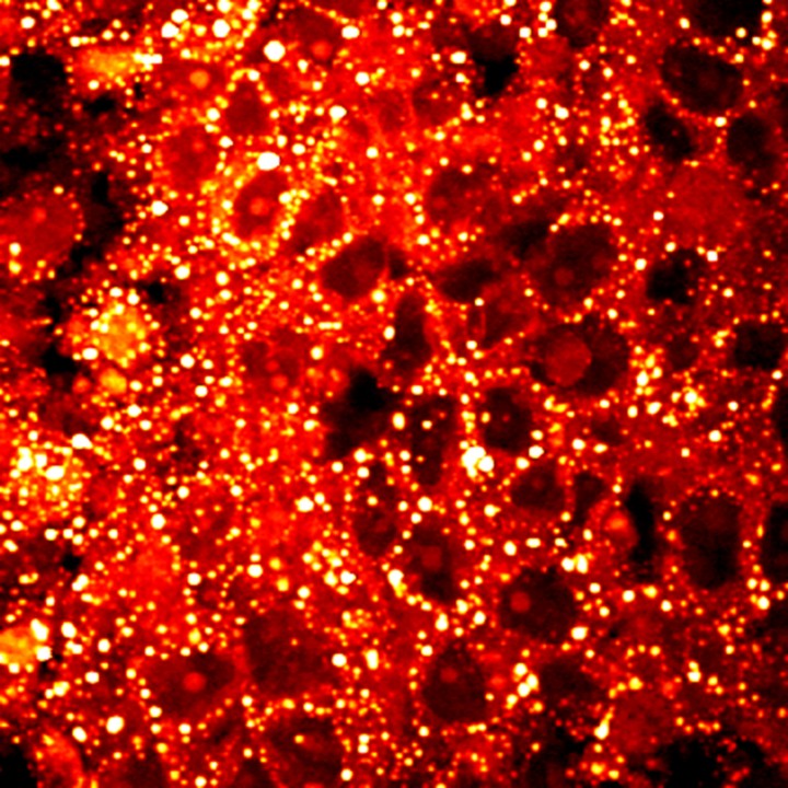
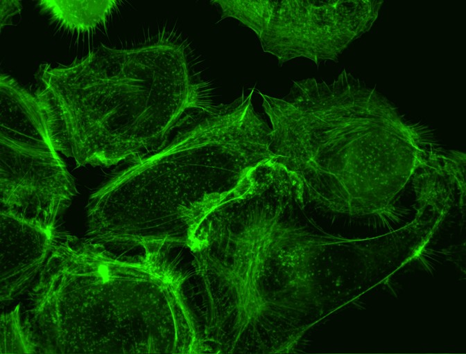
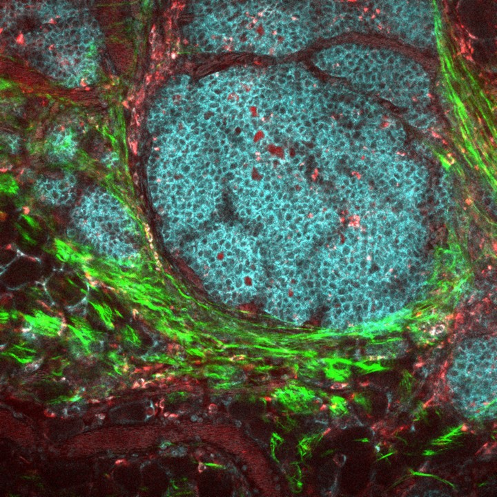
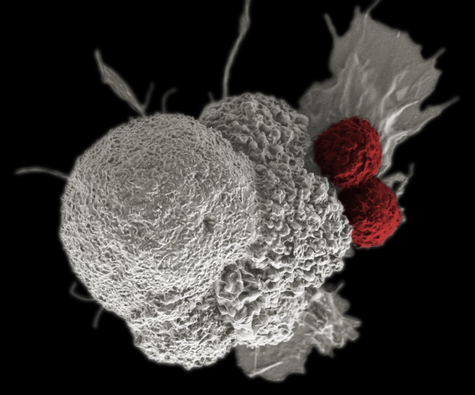
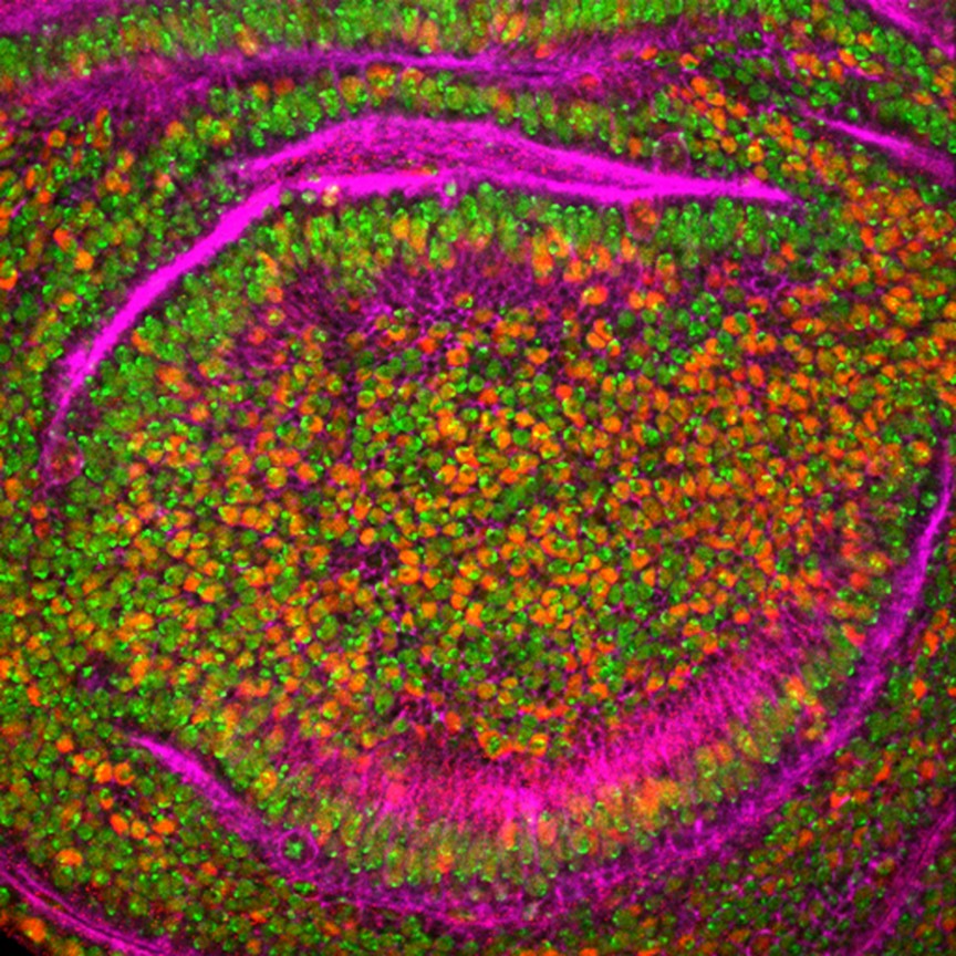
Sometimes, nature is an abstract expressionist.

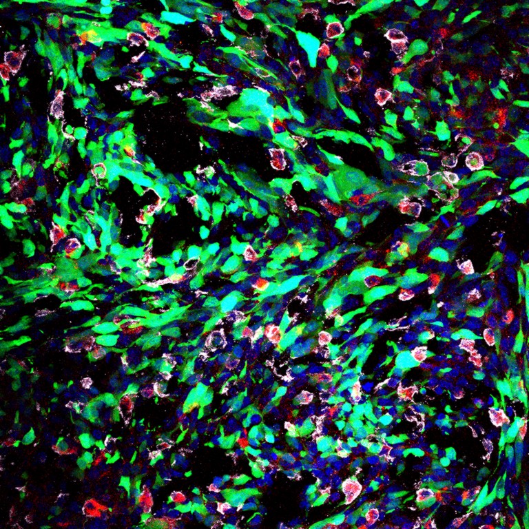
Below are selected images from the National Cancer Institute's "Cancer Close Up 2016" collection, posted last month.

The NCI in February put out a call to researchers for "spectacular microscopy photographs of cancer-related cells, tissues, and molecules that colorfully illustrate significant research conducted by NCI-designated cancer centers."

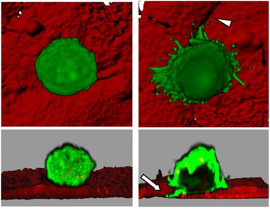
The NCI said it was particularly interested in images related to precision medicine, for which the agency receives funding as part of the federal government's Precision Medicine Initiative.

Most of the images were created using fluorescence microscope, an NCI spokesperson said. Some used an electron microscope. The images will adorn its exhibit booth at annual cancer research meetings and will be featured in public image galleries (not to mention Instagram).

Sponsored

You can see the full collection here, but we liked these the best. The descriptions are from the NCI and have been lightly edited.

ncilipid
Altered lipid metabolism may be a key signature of cancer. This image reveals the storage of cholesterol ester in lipid droplets (bright dots) in aggressive human prostate cancer. This finding may lead to new cancer drugs that work by blocking the activity of cholesterol esterification. (Ji-Xin Cheng/NCI)


ncioncoprotein
Infection with certain types of human papillomavirus (HPV) is associated with various cancers. Researchers are working to understand the processes by which HPV can transform a healthy cell into a cancerous one. This image shows actin (stained in green), a protein involved in cell motion, in HPV-16 E6 and E7-expressing human foreskin keratinocytes. (Ewa Krawczyk/NCI)


ncibreasttumor
This image of a breast cancer tumor and its microenvironment was obtained from a live mouse model. The image was obtained without any fluorophores, stains, or dyes, using only the metabolic co-factors of NADH and FAD, which are already inside of cells, along with second harmonic generation to see collagen. This technique has important clinical potential for patients who require label-free imaging, and may lead to more effective diagnoses and treatments. Tumor cells display in cyan, macrophages in red, collagen fibers in green. (Joseph Szulczewski, David Inman, Kevin Eliceiri and Patricia Keely/NCI)


nciimmunotherapy
Shown here is a pseudo-colored scanning electron micrograph of an oral squamous cancer cell (white) being attacked by two cytotoxic T cells (red), part of a natural immune response. Nanomedicine researchers are creating personalized cancer vaccines by loading neoantigens identified from the patient's tumor into nanoparticles. When presented with immune stimulants, this activates the patient's own immune system, leading to expansion of tumor-specific cytotoxic T cells. (Rita Elena Serda/NCI)


nciDrosophilia
The human CBFA2T3-GLIS2 fusion protein is a key driver of pediatric acute megakaryoblastic leukemia (AMKL), and confers a poor prognosis. Researchers found a way to express CBFA2T3-GLIS2 (red) in larval fruit fly wing disc cells. This confirms a major role for a pathway that may provide a target for new therapies. Nuclei (green) and actin filaments (purple) are also shown. (Suresh Marada/NCI)


nci nanoparticles
Treating cancer in the brain is particularly difficult because most drug molecules are not small enough to penetrate the blood-brain barrier. Researchers wonder whether nanoparticles can serve as a drug-delivery mechanism. This image shows nanoparticles (red) being taken up in the brain of a live rat model with glioblastoma (in green). Nuclear DNA is in blue; tumor-associated macrophages in white. (Eric Hoyeon Song, Alice Gaudin, W. Mark Saltzman/NCI)

More detail on how the images were created from The Washington Post:

Most of the photos ... are made with fluorescent optical microscopes with attached digital cameras. Scientists use various fluorescent stains to highlight specific cellular structures ... and these stains and dyes “light up” when exposed to specific wavelengths of light. (Often) the final multicolored images obtained are composites — overlays — of separate images taken using different wavelengths.

And from STAT:

The photographic techniques that produced them are allowing scientists to understand how the interactions between cellular proteins and the cell nucleus allow cancer cells to invade far-flung tissue, for instance, and how certain molecules allow tumor cells to withstand chemotherapy.

lower waypoint
next waypoint
Homeowners Insurance Market Stretched Even Thinner as 2 More Companies Leave CaliforniaAs California Seeks to Legalize Psychedelics for Therapy, Oregon Provides Key LessonsWatch Ferns Get FreakyCalifornia’s Commercial Salmon Season Is Closed Again This YearHoping for a 2024 'Super Bloom'? Where to See Wildflowers in the Bay AreaWhere to See Cherry Blossoms in the Bay Area This SpringThese Face Mites Really Grow on YouIs It Time for an Essential California Energy Code to Get a Climate Edit?Love Is an Albatross. Literally. Watch These Birds Do a Courtship DanceInsurance In California Is Changing. Here's How It May Affect You